How a Brain CT Scan Reveals the Source of Cognitive Health Problems
Introduction
Have you or a loved one ever needed a brain scan after a sudden headache, a bad fall, or confusion that came on fast? If so, you are not alone.

A brain CT scan, also called a head CT or CAT scan, is often the very first test doctors use when they need fast answers about what is happening inside your head.
A brain CT uses special X-ray equipment to create detailed cross-sectional images of your skull and brain. According to the Mayo Clinic’s guide on brain CT scans, this test helps assess head injuries, severe headaches, dizziness, and symptoms of stroke or aneurysm. It is quick, painless, and widely available at most hospitals and imaging centers.
For anyone worried about cognitive health, the brain CT is a powerful tool. It delivers rapid anatomical detail that helps doctors spot acute changes and plan treatment. And if you are recovering from a brain injury or other neurological event, repeat scans can track how your brain heals over time. This idea of a "reboot brain" — using imaging to guide recovery — is becoming more common in modern medicine.
In this article, we will explain how brain CT works, what it reveals about your cognitive health, and what to expect before, during, and after the scan. We will also touch on related methods like MRV brain scans, emerging approaches such as magnetic brain stimulation, and the role of preclinical research in shaping future brain health tools.
If you want to understand how your brain function connects to your daily concentration, check out this guide on improve concentration with neuroscience.
For a deeper look at how modern systems affect the way you think, explore this note on the Value Reinforcement System.
What Is a Brain CT Scan?
A brain CT scan, also called a head CT or CAT scan, is a medical imaging test that gives doctors a detailed look inside your skull. It works by taking a series of X-ray pictures from many different angles. A computer then combines those images into cross-sectional slices. Think of it like a loaf of bread where each slice shows a different layer of your brain and bone.
This test is especially useful in emergencies. According to the RadiologyInfo’s head CT scan guide, it helps assess head injuries, severe headaches, dizziness, and symptoms of stroke or aneurysm. It can quickly show bleeding inside the brain, skull fractures, large tumors, or fluid buildup. That speed often helps doctors save precious time.
Modern CT scanners are incredibly precise. They can capture images with sub-millimeter resolution. That means they can spot very small changes that older machines might miss. And you can find these scanners at almost any hospital or imaging center across the country.
It is good to know how brain CT compares to other imaging methods. For example, an MRI uses strong magnets and gives better detail for soft tissue. A specialized MRV brain scan focuses specifically on the veins in your head. CT is often the first choice for trauma because it is fast, widely available, and excellent at detecting fresh bleeding.
Your brain’s structure matters for your daily thinking and focus. If you want to take steps to sharpen your attention, try these training your brain to focus longer strategies.
Brain CT vs. MRI vs. PET: Choosing the Right Imaging Tool
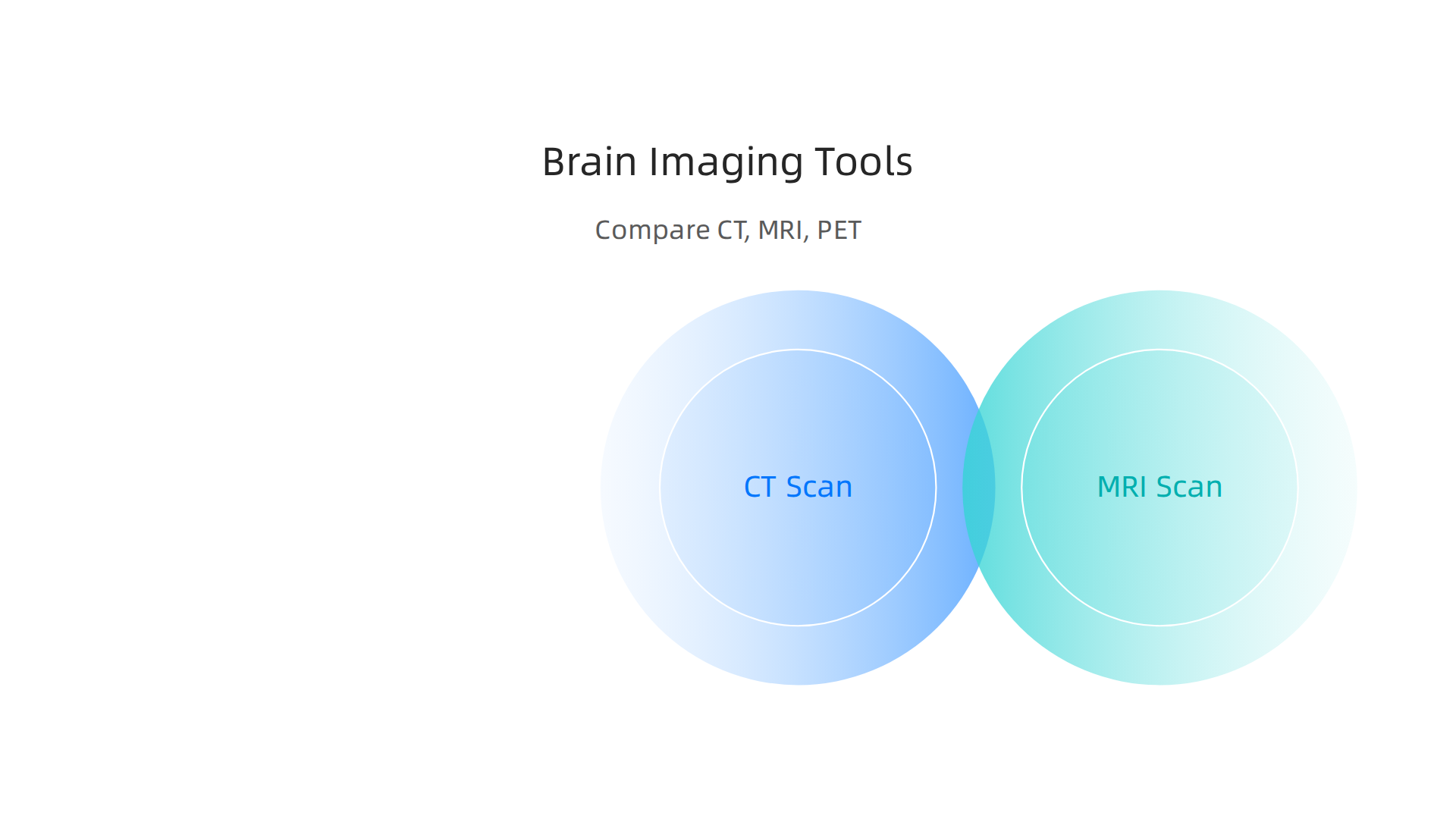
Now that you know what a brain CT scan can do, you might wonder when doctors pick it over other options. The three main brain imaging tools are CT, MRI, and PET. Each one has a special job.
When CT wins the race
A brain CT is the go-to choice in emergencies. It is fast. A scan takes just a few minutes. It is great at spotting bleeding, skull fractures, and large tumors right after an injury or stroke. If someone comes into the ER with a head injury or sudden weakness on one side, CT is usually the first stop. That speed can save a life by helping doctors make quick decisions about treatment.
When MRI gives better detail
MRI uses strong magnets instead of X-rays. It gives much sharper pictures of soft tissue inside your skull. An MRI can spot small white matter lesions, early tumors, and early signs of neurodegenerative disease that CT might miss. According to the ACR Appropriateness Criteria guide on imaging for dementia, MRI without contrast is usually the preferred first test for evaluating cognitive decline. The trade-off is time. An MRI takes 30 to 60 minutes and requires you to lie very still inside a loud tube. For patients who cannot tolerate that, or for trauma situations, CT remains the smarter choice.
When PET adds another layer
PET scans do something different. They do not just show structure. They show activity. A PET scan tracks how your brain uses glucose or oxygen. This tells doctors which parts of your brain are working hard and which parts are slowing down. PET is often used alongside CT for conditions like dementia and epilepsy. It can help tell different types of dementia apart. For example, Alzheimer’s disease and frontotemporal dementia show different patterns on a PET scan.
How doctors decide
So which test is right for you? It depends on what the doctor is looking for. For a suspected stroke or head trauma, brain CT is usually the first choice. For ongoing memory problems or suspected early dementia, MRI is often selected first. For complex cases where structure alone does not tell the full story, PET with CT can give the functional picture doctors need.
Understanding how your brain looks on a scan is one thing. Understanding how your daily habits shape your attention over time is another thing entirely. The VRS Patent 12,205,176 describes a behavioral framework that explains how reinforcement shapes focus and concentration at a system level. It is a different lens for thinking about cognitive health.
If you want to train your brain for better concentration, you can explore neuroscience-backed methods to improve focus that complement what medical imaging reveals about your brain.
When Is a Brain CT Used for Cognitive Health?
You might think a brain CT is only for emergencies like car accidents or sudden strokes. And yes, that is a big part of its job. But a brain CT also plays a surprising role in cognitive health. For people dealing with memory loss, confusion, or other thinking problems, a CT scan often serves as the first step in finding an answer.
The emergency cases are clear.
When someone suddenly cannot speak, loses strength on one side, or has a bad fall, doctors order a brain CT right away. The scan can spot bleeding inside the skull, a blocked blood vessel from a stroke, or a skull fracture within minutes. This speed can be the difference between recovery and long-term damage. For acute intracranial hemorrhage and traumatic brain injury, CT is the gold standard.
The less obvious cases matter just as much.
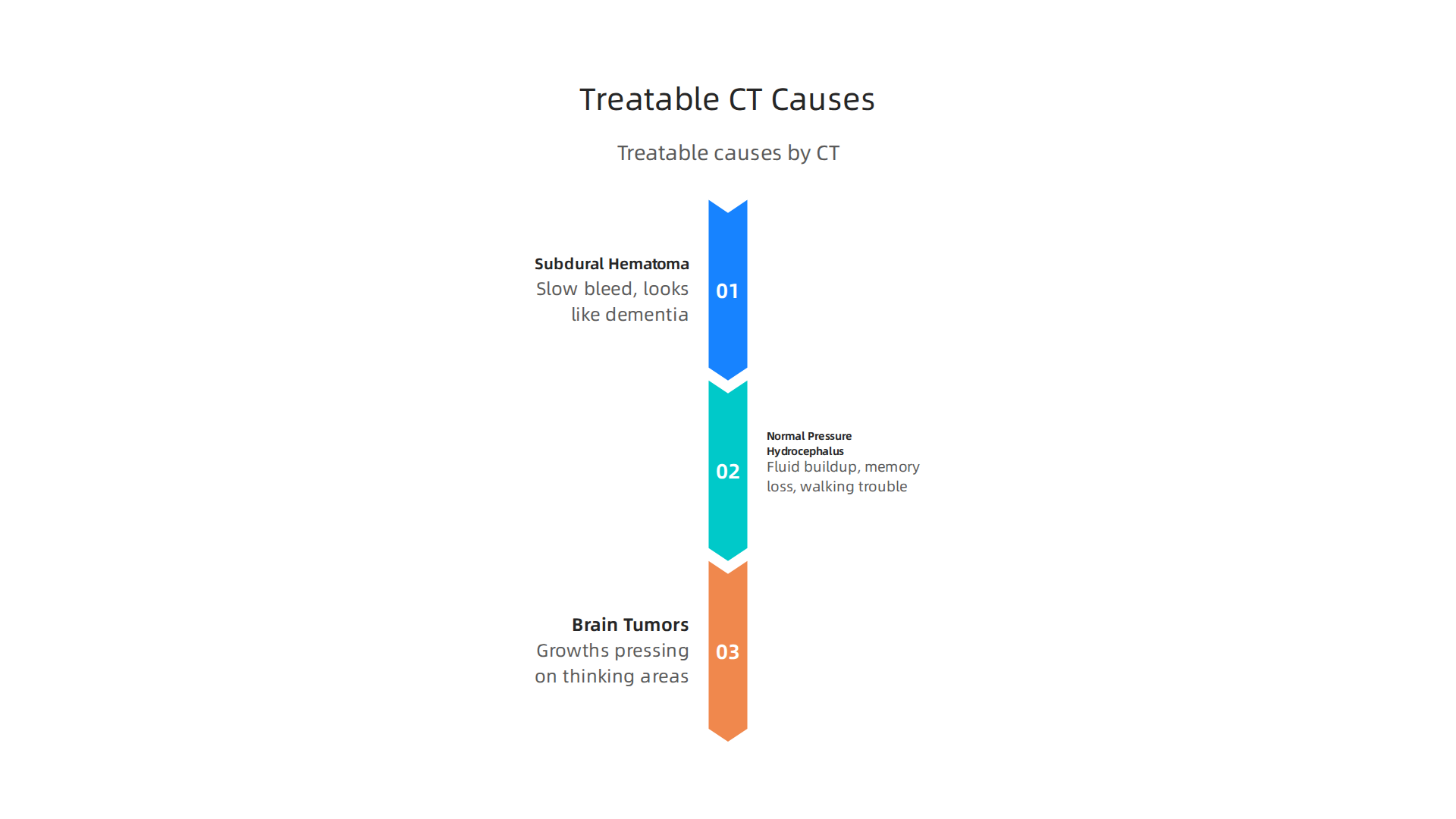
Here is something many people do not realize. Some causes of chronic cognitive decline are actually treatable. A brain CT can find them. For example, a subdural hematoma is a slow bleed on the brain’s surface that can happen after a minor head injury. In older adults, this can look just like dementia. Someone might seem confused, forgetful, or withdrawn. But a simple CT scan can spot the blood, and surgery can fix the problem.
The same goes for normal pressure hydrocephalus, a condition where fluid builds up in the brain. It causes trouble walking, memory loss, and losing control of urine. A CT scan shows the enlarged fluid spaces clearly. Once diagnosed, a shunt can drain the fluid and reverse many symptoms. Brain tumors that press on thinking areas can also be found this way.
What guidelines say.
The ACR Appropriateness Criteria for dementia imaging 2024 update notes that CT without contrast is usually appropriate for initial imaging in patients with cognitive decline. If you have new headaches along with thinking changes or a focal neurological deficit like weakness in one arm or leg, CT is often the recommended first test.
The bottom line is this. If you or someone you know has sudden cognitive changes or a slow decline that worries you, a brain CT might be the right place to start. It is fast, widely available, and can find problems that are fixable.
When imaging gives doctors a clear picture of what is happening in your brain, they can make better decisions about what to do next. And for those looking to sharpen their focus after a health scare or as part of everyday wellness, there are practical ways to train your brain to focus longer that build on what your scan reveals.
Understanding the structure of your brain is one piece of the puzzle. Understanding how your daily habits shape your thinking patterns is another. A Recognition Systems note on the Value Reinforcement System explores how the modern world changes the way we pay attention, giving you a bigger view of cognitive health.
What to Expect During a Brain CT Scan
If your doctor orders a brain CT, you might feel a little unsure about what happens next. Don’t worry. The test is simple, painless, and very fast.
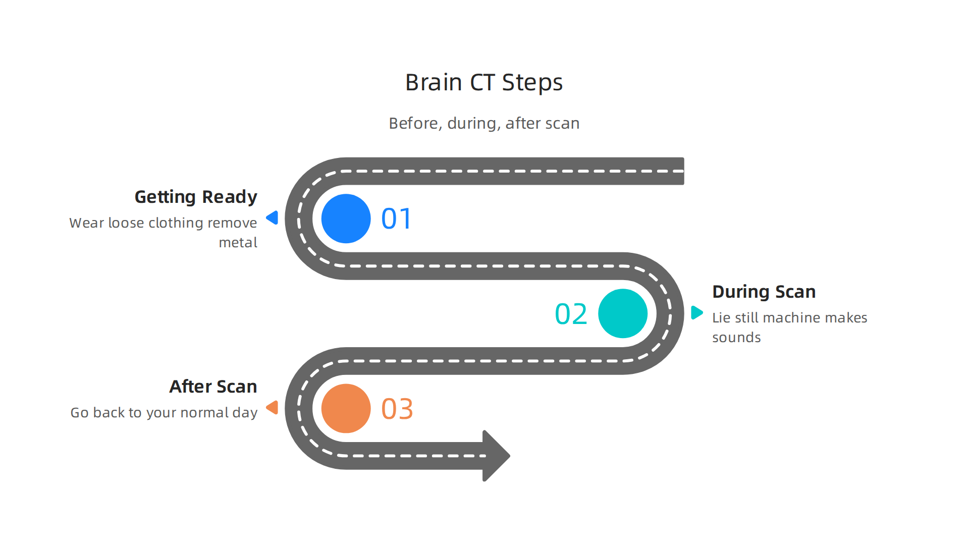

Most scans are done in about 5 to 10 minutes.
Getting ready for the scan.
In most cases, you don’t need to do anything special to prepare. You can eat, drink, and take your regular medications. The Mayo Clinic recommends you wear comfortable, loose-fitting clothing to your appointment. You will also need to remove any metal objects like jewelry, glasses, or hearing aids.
If your doctor wants to use contrast dye, the preparation changes a bit. Contrast helps blood vessels and certain brain problems show up more clearly. You might be asked not to eat or drink for a few hours before the scan. You will also need a quick check of your kidney function because your kidneys remove the dye from your body. Let your doctor know if you have any history of allergies.
What happens during the scan.
When it is time for the test, you lie flat on your back on a table. A technologist will position you and may use a soft strap to help keep your head still. According to Radiologyinfo.org, the table slides into the round opening of the scanner. The X-ray tube rotates around your head taking images.
You need to stay very still during the scan. Moving can blur the pictures. The machine makes buzzing and clicking sounds, which is totally normal. The technologist watches you from another room but can talk to you through a speaker. You can talk to them, too.
If you get contrast, you might feel a warm flush spreading through your body or a metallic taste in your mouth. This feeling goes away quickly.
After the scan.
Once the scan is done, you can go right back to your normal day. A radiologist reads the images and sends a report to your doctor.
Knowing what the structure of your brain looks like gives you a strong foundation. It helps rule out serious problems or find something treatable. Once you have that clarity, working on your daily focus habits makes even more sense. You can build on your brain health by trying neuroscience-backed methods for concentration. These strategies help you stay sharp for the long run.
Interpreting Brain CT Findings in Cognitive Health
When your doctor reviews your brain CT results, they look for a mix of normal changes and potential problems.

Understanding what these findings mean helps you take the right next steps for your cognitive health.
Normal changes you might see.
As we age, our brains change in predictable ways. A brain CT often shows mild atrophy, or slight shrinkage of brain tissue. This is expected and usually does not cause major problems. You may also see small white matter hypodensities. These are tiny spots that appear darker on the scan. Most people have some of these as they get older. They are part of normal aging.
Findings that need attention.
A brain CT is excellent at spotting certain problems that can affect your thinking and memory. Here are the most common ones doctors look for:
- Ischemic strokes. Blocked blood flow to brain tissue shows up clearly on a scan.
- Microbleeds. Tiny areas of bleeding that can signal blood vessel disease.
- Tumors. Growths that may press on healthy brain areas and cause symptoms.
- Hydrocephalus. Extra fluid inside the skull that can slow your thinking and walking.
- Subdural hematomas. Blood pooling around the brain, often from a minor fall.
Spotting these issues early makes a real difference in treatment options. If caught soon enough, many of these problems can be managed or reversed.
What CT does not show well.
Here is an important limit you should know. A brain CT is not as good as an MRI at finding very early changes from dementia or Alzheimer’s disease. MRI shows those tiny changes in brain tissue much better.
But that does not mean a brain CT is useless in cognitive health. It plays a key role. It helps rule out reversible causes of memory loss and confusion. According to the Mayo Clinic’s brain CT scan overview, the test is fast and reliable for spotting problems that need urgent care. A slow brain bleed, a tumor growing quietly, or fluid buildup can all cause symptoms that look like dementia. A CT scan finds these problems, and many of them are treatable.
What this means for you.
Your brain CT results give you a clear starting point. If everything looks normal for your age, you can focus on healthy habits to keep your brain sharp. If something shows up, you have a chance to treat it early before it gets worse.
The way radiologists analyze these scans follows careful, reproducible methods. Understanding this process helps you trust the results and ask better questions. For a deeper look at how imaging data workflows and systematic analysis work in practice, check out the white paper on CRISP-DM and Skylab USA.
For the long haul, building daily focus habits supports your cognitive wellness. Many people find that training your brain to focus longer makes a real difference in how sharp they feel day to day.
Risks and Limitations of Brain CT Imaging
While a brain CT scan is a powerful tool, it is not perfect. Knowing the risks and limits helps you make an informed choice about your health.
Radiation exposure is the main concern.
A brain CT uses a small amount of ionizing radiation to create images. For most adults, the dose is low and the benefit far outweighs the risk. But it is still worth understanding. A head CT exposes you to about the same amount of radiation you get from natural sources over several months. To put this in context, the FDA explains in their patient guide on What are the Radiation Risks from CT that the risk of developing cancer from a single CT scan is very small. Modern scanners use dose-reduction techniques to keep exposure as low as possible. Children are more sensitive to radiation, so doctors are extra careful when scanning young patients.
Contrast reactions are rare but real.
Sometimes doctors use a contrast dye during a brain CT to see blood vessels and tissues more clearly. Most people tolerate this dye without problems. But a small number of people have an allergic reaction. Symptoms can range from mild itching to more serious breathing trouble. Another rare risk is contrast-induced nephropathy, which affects the kidneys. Your doctor checks your kidney function before using contrast to keep you safe.
Where brain CT falls short.
A brain CT has two important limits compared to MRI. First, it does not show soft tissues as clearly. MRI captures finer details in brain tissue, which matters for spotting early dementia changes. Second, artifacts can mess up the image. Things like dental fillings, metal implants, or even patient movement can create streaks or blurs on the scan. As a detailed MRI vs CT vs X-Ray comparison points out, MRI uses no radiation at all, which makes it a safer choice for repeat imaging or for younger patients.
The bottom line is simple. The radiation risk is small, contrast reactions are uncommon, and the image limits are well understood. Your doctor weighs all of these factors before ordering a brain CT. Being aware of these risks and limitations helps you have a better conversation with your healthcare team.
If you are looking to protect your brain health beyond scans, building daily focus habits is a smart move. Many people find that training your brain to focus longer makes a real difference in how sharp they feel over time.
The Role of CT in Brain Recovery and Rehabilitation
When a brain injury happens, the first CT scan is often just the beginning. Repeat brain CT scans become a vital tool for monitoring how the brain heals over time.

Tracking dangerous changes after injury
After a head injury or surgery, doctors watch closely for hemorrhagic expansion — bleeding that gets worse. They also check for edema, which is swelling in the brain. A repeat brain CT can show these changes quickly and clearly. This helps the medical team decide if they need to step in with treatment. The radiation dose from each head CT is low, so the small risk is worth the benefit of catching a problem early. According to Radiation Dose from X-Ray and CT Exams, the amount of radiation from a head CT is about the same as what you get from natural background sources over several months. That is well within a safe range for most patients.
Checking blood flow in stroke recovery
For people recovering from a stroke, a different type of CT can be very useful. CT angiography looks at the blood vessels in the brain to see if they are open or blocked. Perfusion CT measures how well blood is flowing to different areas. Together, these scans help doctors decide on treatments like clot removal or blood pressure management. This is a key part of stroke rehabilitation and can guide therapy decisions for weeks after the event.
Guiding surgery and pressure relief
Some brain injuries lead to a buildup of fluid called hydrocephalus. When that happens, doctors need to place a shunt to drain the fluid. A brain CT helps them decide when to put the shunt in and whether it is working later. The same is true for hematoma evacuation — removing a clot that is pressing on the brain. Serial CT scans over time tell the team if the clot is shrinking or if more surgery is needed.
Supporting long-term recovery with smart habits
Recovering from a brain injury takes time and patience. Alongside medical imaging, building good focus habits can help you regain mental clarity. A practical guide on improving concentration with evidence based strategies offers simple steps to sharpen your attention during the recovery process. And if you want to understand the behavioral science behind staying motivated in rehab, The Science of Gamification gives you a peer reviewed look at how rewards and routines can retrain your brain.
Future Directions in Brain CT Technology
The tools used for brain imaging are getting better fast. Here are three big changes coming to brain CT technology that will make scans safer, sharper, and smarter.
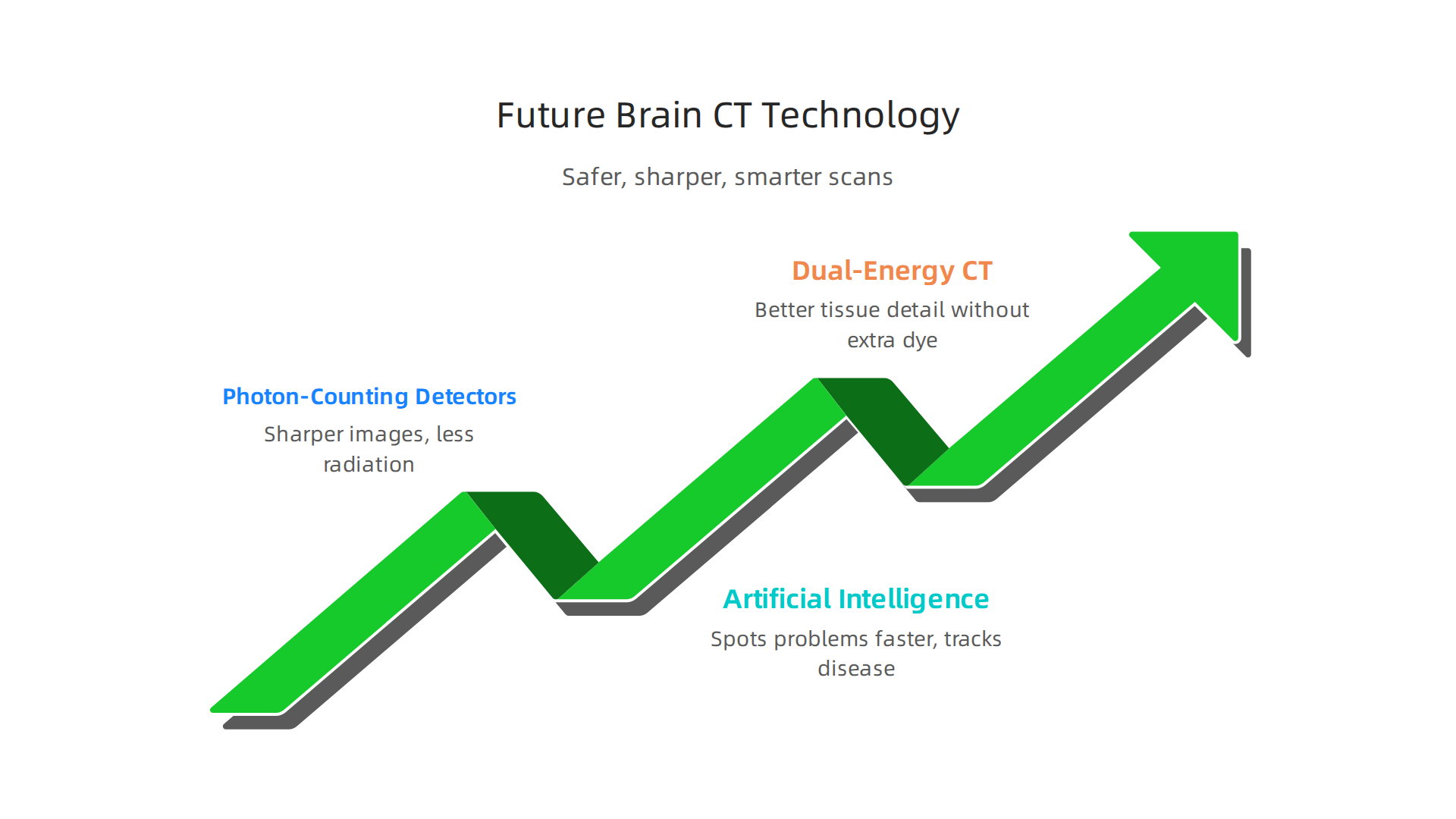
Photon-counting detectors: sharper images, less radiation
Traditional CT scanners measure X-rays in a group. New photon-counting CT detectors count each individual photon instead. This gives doctors much clearer pictures with less noise. It also means they can use a lower dose of radiation. According to a recent article on Photon-Counting CT: High Resolution, Less Radiation, this technology provides similar or better image quality while cutting down on radiation exposure. That is a win for anyone who needs repeat brain scans. Hospitals are already starting to adopt these scanners, and experts at Mass General Brigham are leading the way with emerging CT technology to advance clinical care.
Artificial intelligence that spots problems faster
Doctors are good at reading brain CT scans, but they are human. Artificial intelligence can help by automatically detecting signs of bleeding, stroke, or brain atrophy right away. AI tools can flag urgent findings in seconds, which speeds up treatment decisions. They can also measure how much brain tissue has shrunk over time, helping doctors track diseases like dementia more precisely. This kind of smart assistance will make brain CT even more reliable in the years ahead. One interesting angle to watch is how companies like Meta are exploring simulation-based approaches to protect brain data. A recent report on a Meta patent contrast shows how big tech is thinking about cognitive privacy alongside imaging advances.
Dual-energy CT: better tissue detail without extra dye
Standard CT uses one energy level. Dual-energy CT uses two, which lets it tell apart different types of tissue more clearly. In some cases, this reduces or even removes the need for contrast dye injection. For patients with kidney concerns or allergies, that is a big deal. It also makes it easier to spot small tumors or bleeding that might otherwise be missed.
These innovations mean the future of brain CT is brighter, safer, and more helpful for patients and doctors alike.
Summary
A brain CT (head CT) is a fast X‑ray based scan that gives doctors cross‑sectional images of the skull and brain, making it the first-line test in emergencies like head trauma or suspected stroke and a useful initial tool for certain cognitive problems. This article explains how CT works, when clinicians prefer CT over MRI or PET, and why CT can quickly identify treatable causes of confusion such as subdural hematoma, hydrocephalus, or large tumors. You’ll learn what to expect before, during, and after a scan, how common CT findings are interpreted, and the main risks—especially radiation exposure and rare contrast reactions. The piece also covers how serial CTs guide recovery after injury and highlights future improvements like photon‑counting detectors, AI interpretation, and dual‑energy scans. After reading, you should know when a CT is appropriate, how to prepare, what results mean for cognitive health, and how imaging fits into longer‑term brain recovery and attention‑building strategies.